


Context
Direct electrical stimulation of the brain (DES) has been used for almost a century as a tool for intraoperative functional exploration in an awake patient (Penfield 1937, 1947). The principle is as follows: by transiently applying a focal electrical current, it is observed that the functioning of the patient is transiently disturbed. This tool allows real-time mapping of essential functional regions (language, motor skills) and is nowadays used successfully in clinical practice in the awake surgery of epileptogenic foci and brain tumors: resection can be pushed safely up to functional boundaries.
It is remarkable that, despite its proven clinical efficiency at the behavourial level, very little is known about the electrophysiological effects of electrical stimulation.
The understanding of these effects, beyond fundamental neuroscience interests, is also crucial for the emerging field of new technological neurodevices that requires the development and validation of improved stimulation devices and protocols for acute use (awake surgery) and chronic use (therapeutic neuromodulation and neuroprostheses that could allow a sensorimotor restoration notably through brain-machine interfaces). At an applied and clinical level, we have also planned to use the electrophysiology evoked by the direct electrical stimulation (DES) of the brain during brain surgery to diagnose and determine the anatomical connectivity on-line in order to guide the surgery in patients. This need to go beyond the proof of concept we have alrealdy performed and necessitate to address and solve some methodological challenges.
Three different types of evoked potentials should can be recorded with different methodologies:
– Direct cortical response (DCR), when recording the cortex in the immediate vicinity of the stimulation site,
– Cortico-(Axono)-Cortical Evoked Potentials: (CCEP or sometimes CACEP), i.e. recording the cortex at a distant site from the stimulating site. These potentials are elicited by physiological propagation through white matter associative and striato-thalamo-cortical pathways from the locally stimulated area towards the distal area,
– Axono-cortical evoked potentials (ACEP), when the cortex is distally recorded from a stimulation site within the white matter.
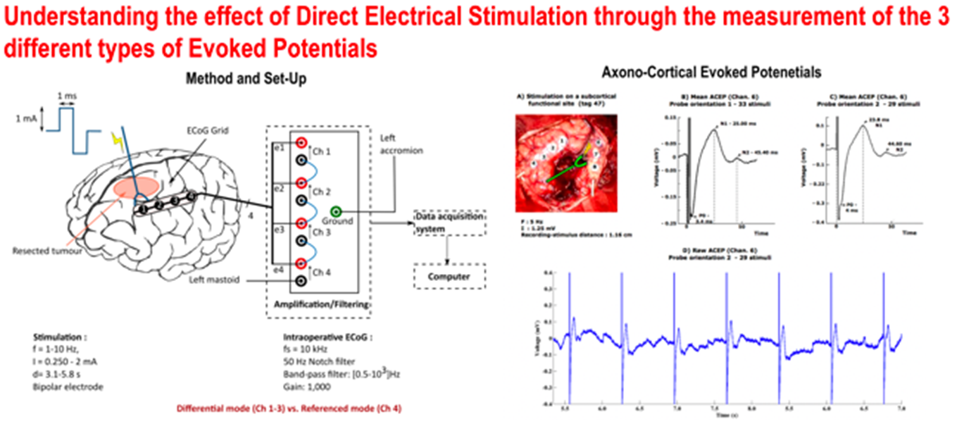 Figure 2. Basic Method and Set-up to record potentials evoked by DES with an example of recorded ACEP.
These evoked potentials are technically difficult to observe, particularly in the operating theater in which the environment is noisy. Their registration requires careful methodological considerations as to how they can be measured. To improve our knowledge of the effects of DES and to generalize the approach of electrophysiological mapping by evoked potentials we intend to scan the effect of different stimulation parameters by combining the two different methodologies.
Different manipulations of the stimulation parameters will be performed: intensity, pulse width, inter-electrode distance and eventually frequency. For instance, it is expected that the variations of these parameters could facilitate the appearance of remote CCEP which indicate the connectivity between the stimulation and the recording sites. Some of these testing have been already performed by the Camin team in patients operated in awake surgery (see publications). However the surface covered by the ECoG grids used for recording the EP is quite small. By contrast, the ECoG grids used in epileptic patients is much larger and the intensity can be higher. A combinaison between a flexible stimutator as that use in awake surgery (France) and the methodology used for recordings of EP in epileptic patients (Japan) will allow to scan the effect of different parameters efficiently.